
Picking an E3 Ligase Assay: FP, TR-FRET, ELISA, NanoBRET and More
Most degrader programs still screen degradation-first: run a cellular assay, see whether the target disappears, and iterate. The problem is that a compound can fail for several independent reasons, including weak E3 engagement, poor permeability, unproductive ternary geometry, inefficient ubiquitination, and a degradation readout cannot tell them apart. A binding assay isolates one variable: how tightly the compound engages the ligase, with no cells, no permeability, no proteasome in the way. Choosing an E3 ligase binding assay platform is where a program gets its first interpretable number. The human genome encodes over 600 E3 ubiquitin ligases, but the working E3 ligase list remains dominated by CRBN and VHL, with DCAF1, DCAF11, DCAF16, MDM2, IAP, and KEAP1 gaining ground. Whichever ligase you recruit, the same decision tree applies.
Comparing E3 Ligase Assay Technologies
These platforms detect different physical events (rotational mobility, proximity, heat, refractive index) and differ mostly in what each measurement costs you.
- Fluorescence polarization (FP): binding assay. A tracer tumbles fast when free and slowly when bound, so polarization reports occupancy. In competitive binding assays an unlabeled compound displaces it, and the IC50 converts to Ki. Only the tracer is labeled, nothing is immobilized, and there are no washes, so the fluorescence polarization assay runs in minutes on a standard fluorescence plate reader.
- Time-resolved FRET (TR-FRET): binding assay. A TR-FRET binding assay reads a lanthanide donor after a time delay, so background fluorescence has decayed before acquisition; time-resolved fluorescence also reaches ternary complexes directly. Both partners need labeling, the readout is ratiometric, and quenchers hitting one channel distort it.
- Amplified luminescent proximity homogeneous assay (ALPHA) — binding assat. Bead proximity chemiluminescence with excellent signal-to-background, though the chemistry is quenchable and donor beads are light-sensitive.
- Isothermal titration calorimetry (ITC), surface plasmon resonance (SPR) and bio-layer interferometry (BLI): binding assays, low throughput. ITC returns Kd, stoichiometry and thermodynamics label-free in one 40-minute titration on ~200 µL; SPR and BLI add kinetics, at the cost of immobilization and costly sensors.
- NanoBRET assay and in-cell ELISA: functional assays. The nano-bioluminescence resonance energy transfer (NanoBRET) assay follows engagement and ternary complex formation in live cells, and a NanoBRET ubiquitination assay reads ubiquitin transfer, but both need tagged constructs. An enzyme-linked immunosorbent assay (ELISA) kit quantifies target protein in fixed cells; ELISA assay results rank ligands by how well they rescue it from a reference degrader. Both report outcomes, not occupancy.
Cell-Free vs. Cell-Based Formats: What Each Platform Actually Measures
- Cell-free formats use purified protein. They measure binding: the binary affinity of a compound for the ligase and the ternary complex. That is, the assembly of the protein of interest (POI), the degrader, and the E3 ligase that positions the substrate for ubiquitin transfer. Because no cellular process intervenes, the value is directly attributable, which makes binary affinity the first experimentally accessible checkpoint along the degrader mechanism of action and the earliest point at which a liability can be identified, before resources are committed to cellular studies.
- Cell-based formats use intact cells. They measure function: target ubiquitination and loss of target protein. Here, a weak result can reflect affinity, but equally permeability, metabolic stability, intracellular retention, ligase expression, unproductive ternary geometry or inefficient ubiquitination, and the assay cannot discriminate among them.
This is the practical distinction between a binding assay vs functional assay: cell-free formats identify the cause of a result; cell-based formats establish whether the compound works. Binary affinity is no surrogate for degradation, and the relationship is not monotonic, since excessively tight ligase binding saturates the degrader–ligase species early and narrows the window before a hook effect appears. In the CRBN panel profiled by González-Pico et al., potent cellular degraders generally sat below roughly 500 nM: not a universal threshold, but the kind of guardrail only a ligand binding assay provides.
Throughput, Cost, and Setup Time: Matching the Assay to Your Screening Stage
FP is homogeneous, requiring no washing or separation, so it moves readily into 384-well plates and supports a genuine high-throughput screening assay. Miniaturization is where the economics change: at 20 µL per well instead of 100 µL, roughly four times more compounds can be profiled with the same total reagent, and in CRBN and VHL assays this does not compromise performance, with Ki values in 384-well format matching the 96-well data. That makes FP the natural fit for the analog series stage, where most of the volume sits. Controls still matter: keep the tracer near its Kd, confirm a Z′ factor of at least 0.5, and monitor total fluorescence to flag autofluorescence or precipitation.
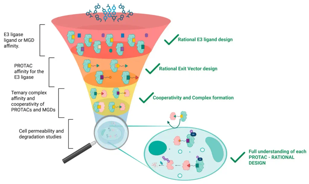
Figure 1. Binding-first workflow for rational degrader discovery. Source: González-Pico L, Camino T, Ortigueira S, Prieto-Díaz R, Sotelo E, Majellaro M. Bridging E3 ligase binding and targeted degradation through fluorescence polarization. Int J Mol Sci. 2026;27(18):8054.
Ternary measurements are a different exercise: saturating the degrader with one protein and titrating the other consumes considerably more purified protein, which is why a TR-FRET assay is often chosen for the ternary curve, while ITC and SPR serve as orthogonal confirmation on a few leads. FP formats for ternary cooperativity are in development.
Setup time deserves equal weight. Building a ligand binding assay by FP from scratch means screening candidate tracers across exit vectors, linkers, and fluorophores, then characterizing photophysics, stability, and Kd before a single compound is measured. That is weeks of development preceding any structure–activity relationship (SAR) data. The same displacement principle underpinned the radioligand binding assay decades earlier, now largely replaced by fluorescence and its freedom from radioactive waste.
Where Ready-to-Use FP Kits Fit Alongside DIY Protocols and Outsourced Panels
Between building everything in-house and outsourcing everything sits a practical middle path. Our ready-to-use E3 ligase kits for VHL and CRBN are built on the tracers and protocols validated against molecular glues, CELMoDs, and clinical-stage proteolysis-targeting chimeras (PROTACs) including vepdegestrant and CFT1946, with 15-minute incubations and standardized formats designed for reproducible measurements across laboratories.
When capacity is the constraint, or when resistance data prompt a pivot between ligases, our FP-based screening services for CRBN and VHL deliver single-point affinity screens or full displacement curves ready to feed your next design cycle.
Deciding which format fits your current stage? Get in touch and we will help you match the assay to the decision you actually need to make!
References
González-Pico L, Camino T, Ortigueira S, Prieto-Díaz R, Sotelo E, Majellaro M. Bridging E3 ligase binding and targeted degradation through fluorescence polarization. Int J Mol Sci. 2026;27(18):8054. doi: 10.3390/ijms27188054
Liu X, Zhang X, Lv D, Yuan Y, Zheng G, Zhou D. Assays and technologies for developing proteolysis targeting chimera degraders. Future Med Chem. 2020;12(12):1155–1179. doi: 10.4155/fmc-2020-0073
Yang K, Zhao Y, Nie X, Wu H, Wang B, Almodovar-Rivera CM, Xie H, Tang W. A cell-based target engagement assay for the identification of cereblon E3 ubiquitin ligase ligands and their application in HDAC6 degraders. Cell Chem Biol. 2020;27(7):866–876. doi: 10.1016/j.chembiol.2020.04.008
Yin L, Shu P, Peng X. Context matters: E3 ligase–ligand pairing strategies for optimized PROTAC performance. Protein Cell. 2026;17(5):384–398. doi: 10.1093/procel/pwaf107
Wurz RP, Rui H, Dellamaggiore K, et al. Affinity and cooperativity modulate ternary complex formation to drive targeted protein degradation. Nat Commun. 2023;14:4177. doi: 10.1038/s41467-023-39434-0
Hall MD, Yasgar A, Peryea T, Braisted JC, Jadhav A, Simeonov A, Coussens NP. Fluorescence polarization assays in high-throughput screening and drug discovery: a review. Methods Appl Fluoresc. 2016;4(2):022001. doi: 10.1088/2050-6120/4/2/022001
Related posts

PROTAC Hook Effect: How E3 Ligase and Target Protein Binding Affinity Shapes Degradation Efficiency

VHL, CRBN, and Emerging Ligases like DCAF: Selecting E3 Ligase Handles for PROTAC Drug Discovery


Fluorescence Polarization Binding Assays for E3 Ligases in Targeted Protein Degradation (TPD) Programs




PERTE SALUD Proyecto Nº: IDI-2025-0751
Título: TERAPIAS AVANZADAS EN INMUNOONCOLOGÍA: UNA ESTRATEGIA INNOVADORA
BASADA EN LA DEGRADACIÓN DIRIGIDA DE GPCR EN EL EJE ADENOSINÉRGICOS

EIC Pathfinder; Project name: Unisens; Universal GPCR Activity Sensor for Next Generation Drug Discovery


Esta entidad fue beneficiaria de las ayudas para la ejecución de acciones de promoción exterior de las empresas gallegas. El objetivo principal de estas ayudas es incentivar la realización de acciones de promoción exterior generadoras de ventajas competitivas. El resultado que se pretende conseguir es el impulso de las pymes y sus productos y servicios, aumentando el número de empresas de base exportadora.
Celtarys Research is beneficiary of a WomenTechEU grant supporting deep-tech start-ups led by women


Principia 2021 Program
Celtarys staff has been funded by the Xunta de Galicia within the framework of the PRINCIPIA 2021 Program of the Galician Innovation Agency (GAIN)
InnovaPEME Program
Celtarys Innovation Plan has been funded by the InnovaPEME 2022 program of the Galician Innovation Agency (GAIN)

Bonos de Innovación de Celtarys Research (026)
Para promover o desenvolvemento tecnolóxico, a innovación e unha investigación de calidade. Esta operación está financiada pola Xunta de Galicia, a través de axudas concedidas pola Axencia Galega de Innovación, dentro do programa de axudas a empresa Bonos de innovación 2022.


Celtarys Research is part of the public-private consortium developing the project PREDICTEAM
New prognostic quantitative biomarker assays for predicting patient response to immune checkpoint inhibitor (ICI) treatment


NEOTEC · CDTI — Ministerio de Ciencia e Innovación
The Project ‘New chemical conjugation technology for therapeutic targets’ has been founded by CDTI under its program NEOTEC. The NEOTEC program objective is supporting the establishment and consolidation of technology-based enterprises. A technology-based company (EBT) is a company whose activity focuses on the exploitation of products or services that require the use of technologies and knowledge developed since the research activity. The EBT base their business strategy or activity in the intensive domain of scientific and technical knowledge. Budget awarded: 416.177€ | Execution dates: 1/1/2023 – 31/03/2024