
VHL, CRBN, and Emerging Ligases like DCAF: Selecting E3 Ligase Handles for PROTAC Drug Discovery
The choice of E3 ligase recruiter is one of the most consequential early decisions in any PROTAC drug discovery program. A PROTAC simultaneously engages a target protein and an E3 ubiquitin ligase, triggering ubiquitination and proteasomal degradation. Tissue expression, ternary complex formation, and resistance liability (for example, through loss of ligase expression in treated tumors) all determine whether a PROTAC makes it from the lab to the clinic. Choosing between VHL, CRBN, and emerging E3 ligase alternatives such as the DCAF family is therefore as strategically important as the design of the target protein ligand itself.
VHL and CRBN: the established E3 ligase handles in PROTAC drug discovery
Von Hippel-Lindau (VHL) and cereblon (CRBN) have dominated targeted protein degradation (TPD) for a straightforward reason: both have well-characterized, ligandable binding pockets with potent small-molecule binders available, and both have been structurally characterized in ternary complexes across diverse target proteins, but there are some differences between them.
While CRBN accommodates thalidomide derivatives through a deep hydrophobic pocket with high conformational plasticity, which underlies the broad substrate scope of CRBN PROTACs, VHL uses a rigid hydroxyproline-recognition motif that generally improves selectivity but restricts neo-substrate diversity. According to recent research, VHL-based PROTACs are much less prone to molecular glue-like behavior, and the higher essentiality of VHL also reduces the potential for fast resistance development compared to CRBN.
Tissue distribution adds another layer: CRBN is broadly expressed with particularly high levels in hematopoietic tissues, explaining its clinical success in hematologic malignancies. VHL is enriched in the kidney, lung, and colon, but expressed at low levels in the liver and CNS, with its stability oxygen-regulated (a concern in hypoxic tumor cores).
Programs should weigh the following when selecting between them:
- Target tissue match: CRBN for hematologic indications; VHL for renal or lung contexts, with caveats around hypoxia.
- Selectivity requirements: VHL’s rigidity tends to produce more selective degraders; CRBN’s plasticity broadens the substrate landscape but increases off-target risk.
- Resistance liability: CRBN is non-essential in many cancer cell lines and is frequently downregulated or mutated in IMiD-refractory patients; VHL loss is a defining feature of clear cell renal carcinoma.

Figure 1. Targeting E3 ligases. Source: Rodríguez-Gimeno A, Galdeano C. Drug Discovery Approaches to Target E3 Ligases. Chembiochem. 2025 Jan 2;26(1):e202400656
Expanding beyond VHL and CRBN: emerging E3 ligases like the DCAF family in TPD programs
DCAF1 is a WD40-domain-containing E3 ligase receptor of the CRL4 complex distinguished by a key property: it is an essential gene. Analysis of genome-wide CRISPR knockout dependency scores shows that DCAF1 essentiality is comparable to DDB1, its direct binding partner, which could limit resistance driven by ligase loss-of-function. This supposes a meaningful advantage over CRBN, which is frequently downregulated in resistant tumors.
A seminal demonstration came from the DCAF1-BTK PROTAC DBt-10, which achieved efficient BTK degradation in cells with acquired resistance to CRBN-BTK PROTACs due to loss of CRBN expression, while showing no loss of antiproliferative activity relative to wild-type cells. A DCAF1-BRD9 PROTAC also achieved effective BRD9 degradation in VHL-null renal carcinoma cells, confirming DCAF as a viable alternative in VHL-deficient contexts.
Beyond DCAF1, the field is actively exploring E3 ligase ligands across both other DCAF subfamily members (including DCAF16 and DCAF11) and structurally unrelated ligases such as KEAP1 and MDM2. The guiding principle to select E3 ligase for PROTACs is E3-tissue matching: aligning the expression profile and structural constraints of the chosen ligase with the disease biology and target localization of the program. No single ligase fits all contexts.
Fluorescence-based binding assays for E3 ligase handle selection and validation
An E3 ligase handle is the small-molecule ligand that engages the ligase and serves as one arm of the PROTAC. Validating that handle (confirming its binding affinity, selectivity, and compatibility with ternary complex formation) is a prerequisite before investing in full PROTAC synthesis and cellular degradation studies.
Fluorescence polarization (FP) E3 ligase assay formats are well-suited to this purpose. FP detects binding through the change in rotational mobility of a small fluorescent tracer upon engaging a much larger protein. When the tracer is pre-bound to the E3 ligase, an unlabeled competitor displaces it in proportion to its affinity. This competitive format produces IC50 values convertible to Ki, requires no washing or separation steps, and scales directly to 384- and 1536-well formats for targeted protein degradation screening.
An E3 ligase ligand validation workflow using FP typically proceeds through three stages:
1- Direct binding to determine the tracer Kd
2- Competitive displacement to rank compound affinities
3- Cooperative binding measurements to assess PROTAC ternary complex formation
Monitoring total fluorescence intensity alongside polarization is essential to flag autofluorescent compounds or quenchers that can generate false positives. A Z′ factor ≥ 0.5 should be confirmed before advancing to High-Throughput-Screening (HTS) campaigns.
For a VHL E3 ligase assay or CRBN ligase binding assay, the labeled tracer is typically a fluorescein- or BODIPY FL-conjugated analog of established VHL and CRBN binders. The fluorescent tag must not occlude the binding interface, and linker length must be optimized to prevent the propeller effect (excess local fluorophore rotation that compresses the assay’s dynamic range).
Ready-to-use E3 ligase kits: accelerating VHL and CRBN assay development
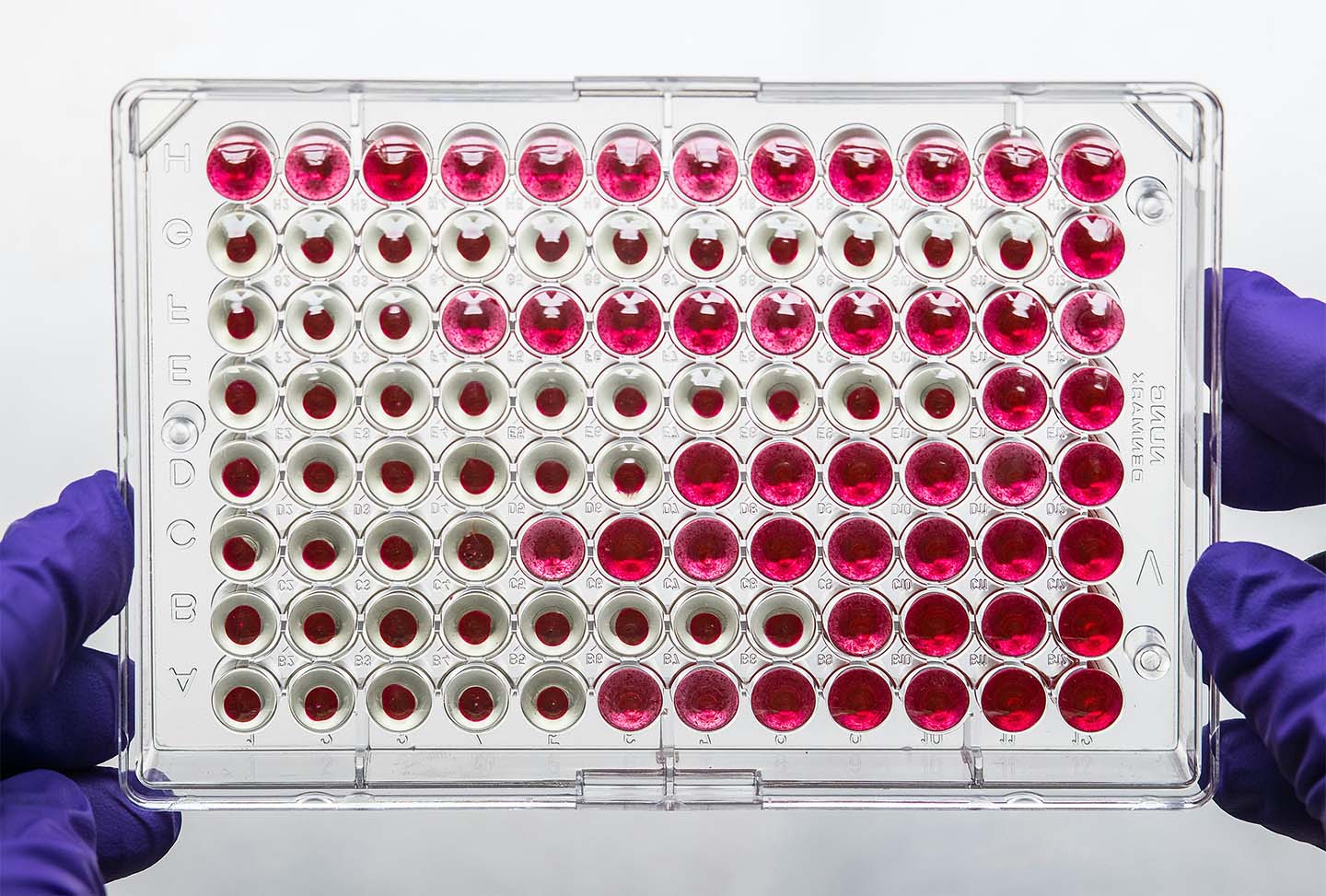
To run a VHL or CRBN E3 ligase binding assay without building validated reagents from scratch, ready-to-use E3 ligase kits designed for fluorescence polarization offer a significant advantage. Pharmacologically validated fluorescent ligands, optimized buffer formulations, and established assay protocols remove the most time-consuming steps from the development cycle, particularly the iterative probe synthesis and tracer Kd characterization described above. An E3 ligase kit for drug discovery format compatible with standard well plate infrastructure can be implemented in-house within a single assay day, with data that directly feeds into PROTAC optimization decisions.
The value of these tools becomes especially clear when programs need to compare E3 ligase handles in parallel, or when resistance data prompt a pivot from one ligase to another, moving quickly without rebuilding the assay from the ground up is a meaningful time saving at the lead optimization stage of PROTAC drug discovery.
More broadly, profiling PROTAC affinity for the E3 ligase before committing to cellular degradation studies supports rational degrader design: binding data help deconvolute the factors governing degradation activity (cell permeability, ternary complex formation, linker geometry, or low ligase expression) that a degradation assay alone cannot resolve.
Developing a PROTAC program demands rigorous biochemical characterization of each E3 ligase handle under consideration, and that characterization starts with the right assay tools. At Celtarys, we offer ready-to-use E3 ligase kits for both VHL and CRBN, including pharmacologically validated fluorescent ligands and optimized FP assay protocols designed for immediate in-house implementation. For programs requiring outsourced profiling, dedicated FP-based screening services deliver affinity binding data and full displacement curves with IC50 and Ki values, ready to inform your next design cycle.
Contact us to discuss how our platform can support your E3 ligase selection and validation workflow!
References
Huang Q et al. Attacking the “undruggable” target: The rise and evolution of targeted protein degradation technologies. Adv Interv Mater. 2026;1:100015. doi: 10.1016/j.advim.2026.100015
Miletić N et al. Workflow for E3 Ligase Ligand Validation for PROTAC Development. ACS Chem Biol. 2025 Feb 21;20(2):507-521. doi: 10.1021/acschembio.4c00812
Osman J, et al. Methods to accelerate PROTAC drug discovery. Biochem J. 2025 Jun 25;482(13):921–37. doi: 10.1042/BCJ20243018
Rodríguez-Gimeno A, Galdeano C. Drug Discovery Approaches to Target E3 Ligases. Chembiochem. 2025 Jan 2;26(1):e202400656. doi: 10.1002/cbic.202400656
Schröder M et al. DCAF1-based PROTACs with activity against clinically validated targets overcoming intrinsic- and acquired-degrader resistance. Nat Commun. 2024 Jan 4;15(1):275. doi: 10.1038/s41467-023-44237-4
Seipp EK, Huang R. Fluorescence polarization binding assays for the E3 ligase FEM1C. Methods Enzymol. 2025;719:347–362. doi: 10.1016/bs.mie.2025.06.002
Related posts
Picking an E3 Ligase Assay: FP, TR-FRET, ELISA, NanoBRET and More

PROTAC Hook Effect: How E3 Ligase and Target Protein Binding Affinity Shapes Degradation Efficiency



Fluorescence Polarization Binding Assays for E3 Ligases in Targeted Protein Degradation (TPD) Programs




PERTE SALUD Proyecto Nº: IDI-2025-0751
Título: TERAPIAS AVANZADAS EN INMUNOONCOLOGÍA: UNA ESTRATEGIA INNOVADORA
BASADA EN LA DEGRADACIÓN DIRIGIDA DE GPCR EN EL EJE ADENOSINÉRGICOS

EIC Pathfinder; Project name: Unisens; Universal GPCR Activity Sensor for Next Generation Drug Discovery


Esta entidad fue beneficiaria de las ayudas para la ejecución de acciones de promoción exterior de las empresas gallegas. El objetivo principal de estas ayudas es incentivar la realización de acciones de promoción exterior generadoras de ventajas competitivas. El resultado que se pretende conseguir es el impulso de las pymes y sus productos y servicios, aumentando el número de empresas de base exportadora.
Celtarys Research is beneficiary of a WomenTechEU grant supporting deep-tech start-ups led by women


Principia 2021 Program
Celtarys staff has been funded by the Xunta de Galicia within the framework of the PRINCIPIA 2021 Program of the Galician Innovation Agency (GAIN)
InnovaPEME Program
Celtarys Innovation Plan has been funded by the InnovaPEME 2022 program of the Galician Innovation Agency (GAIN)

Bonos de Innovación de Celtarys Research (026)
Para promover o desenvolvemento tecnolóxico, a innovación e unha investigación de calidade. Esta operación está financiada pola Xunta de Galicia, a través de axudas concedidas pola Axencia Galega de Innovación, dentro do programa de axudas a empresa Bonos de innovación 2022.


Celtarys Research is part of the public-private consortium developing the project PREDICTEAM
New prognostic quantitative biomarker assays for predicting patient response to immune checkpoint inhibitor (ICI) treatment


NEOTEC · CDTI — Ministerio de Ciencia e Innovación
The Project ‘New chemical conjugation technology for therapeutic targets’ has been founded by CDTI under its program NEOTEC. The NEOTEC program objective is supporting the establishment and consolidation of technology-based enterprises. A technology-based company (EBT) is a company whose activity focuses on the exploitation of products or services that require the use of technologies and knowledge developed since the research activity. The EBT base their business strategy or activity in the intensive domain of scientific and technical knowledge. Budget awarded: 416.177€ | Execution dates: 1/1/2023 – 31/03/2024